Magnetic Resonance - Technology Information Portal Welcome to MRI Technology
Info
  Sheets

Out-
      side


 'pulse sequences' 
SEARCH FOR    
 
  2 3 5 A B C D E F G H I J K L M N O P Q R S T U V W X Y Z
Result : Searchterm 'pulse sequences' found in 1 term [] and 97 definitions []
previous     61 - 65 (of 98)     next
Result Pages : [1]  [2 3 4 5 6 7 8 9 10 11 12 13 14 15 16 17 18 19 20]
Searchterm 'pulse sequences' was also found in the following services: 
spacer
News  (1)  Resources  (5)  Forum  (8)  
 
MRI EquipmentInfoSheet: - Devices -
Intro, 
Types of Magnets, 
Overview, 
etc.
 
The MRI equipment consists of following components:
The magnet generates the magnetic field.
Shim coils make the magnetic field homogeneous.
Radio frequency coils transmit the radio signal into the body part being imaged.
Receiver coils detect the returning radio signals.
Gradient coils provide spatial localization of the signals.
Shielding coils produce a magnetic field that cancels the field from primary coils in regions where it is not desired.
The computer reconstructs the signals into the image.
The MRI scanner room is shielded by a faraday shield.
Different cooling systems cool the magnet, the scanner room and the technique room.

Better MRI equipment and software design along with the latest information technology improves system maintenance and overall communication. Software and digital imaging and communications in medicine (DICOM) compatibility allows to network into hospital databases, helps to modify pulse sequences, data post processing, and archiving via picture archiving and communication system (PACS).

See also the related poll result: 'Most outages of your scanning system are caused by failure of'
Radiology-tip.comradCT Scanner,  Radiography
spacer
Medical-Ultrasound-Imaging.comUltrasound Machine,  Ultrasound System Performance
spacer
 
• Related Searches:
    • Legal Requirements
    • MRI Safety
    • Radio Frequency Coil
    • Magnet
    • Receiver
 
Further Reading:
  News & More:
Low Power MRI Helps Image Lungs, Brings Costs Down
Thursday, 10 October 2019   by www.medgadget.com    
MRI safety targeted as new group offers credentialing test
Monday, 12 January 2015   by www.modernhealthcare.com    
Audio/Video System helps patients relax during MRI scans
Monday, 8 December 2014   by news.thomasnet.com    
Dräger introduces anaesthesia system for MRI environment
Wednesday, 12 December 2007   by www.mtbeurope.info    
MRI Resources 
Process Analysis - Veterinary MRI - Coils - Mobile MRI - Collections - Implant and Prosthesis
 
MRP-7000™InfoSheet: - Devices -
Intro, 
Types of Magnets, 
Overview, 
etc.MRI Resource Directory:
 - Devices -
 
www.hitachimed.com/products/mrp.asp 
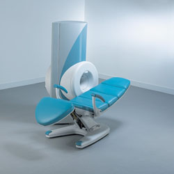
From Hitachi Medical Systems America, Inc.;
because of its dependability, the MRP-7000™ remains popular more than a decade after the first U.S. system was shipped. This system maintains a high resale value, what has made it one of the most sought-after scanners on the used MRI equipment market.
Device Information and Specification
CLINICAL APPLICATION
Whole body
CONFIGURATION
Vertical Orientation Field
DualQuad T/R Body Coil, MA Head, MA C-Spine, MA Shoulder, MA Wrist, MA CTL Spine, MA Knee, MA TMJ, MA Flex Body (3 sizes), Neck, small and large Extremity, PVA (WIP), Breast (WIP), Neurovascular (WIP), Cardiac (WIP) and MA Foot//Ankle (WIP)
SYNCHRONIZATION
Cardiac gating, ECG/peripheral, respiratory gating (2 modes)
PULSE SEQUENCES
SE, GE, GR, IR, FIR, STIR, ss-FSE, FSE, DE-FSE/FIR, FLAIR, ss/ms-EPI, ss/ms EPI- DWI, SSP, MTC, SE/GE-EPI, MRCP, SARGE, RSSG, TRSG, BASG, Angiography: CE, PC, 2D/3D TOF
IMAGING MODES
Single, multislice, volume study
MAGNET TYPE
Permanent, self shielded
STRENGTH
8 mT/m
5-GAUSS FRINGE FIELD
horizontal 2.5 m x 2.1 m vertical
Auto shimming, 3-axis/patient and active shimming
spacer

• View the DATABASE results for 'MRP-7000™' (2).Open this link in a new window

MRI Resources 
Diffusion Weighted Imaging - Non-English - Intraoperative MRI - Calculation - Journals - Guidance
 
MSK-Extreme™InfoSheet: - Devices -
Intro, 
Types of Magnets, 
Overview, 
etc.MRI Resource Directory:
 - Devices -
 
www.onicorp.com/ From ONI Medical Systems, Inc.;
MSK-Extreme™MRI system is a dedicated high field extremity imaging device, designed to provide orthopedic surgeons and other physicians with detailed diagnostic images of the foot, ankle, knee, hand, wrist and elbow, all with the clinical confidence and advantages derived from high field, whole body MRI units. The light weight (less than 650 kg) of the OrthOne System performs rapid patient studies, is easy to operate, has a patient friendly open environment and can be installed in a practice office or hospital, all at a cost similar to a low field extremity machine.
New features include a more powerful operating system that offers increased scan speed as well as a 160-mm knee coil with higher signal to noise ratio, and the option of a CD burner.
Device Information and Specification
CLINICAL APPLICATION
Dedicated extremity imaging
CONFIGURATION
16 cm knee, 18 cm lower extremity;; 12.3 cm upper extremity, additional high resolution v-SPEC Coils: 80 mm, 100 mm, or 145 mm.
SYNCHRONIZATION
No
PULSE SEQUENCES
SE, FSE, GE2D, GE3D, Inversion recovery (IR), Driven Equilibrium, Fat Saturation (FS), STIR, MT, PD, Flow Compensation (FC), RF spoiling, MTE, No Phase Wrap (NPW)
IMAGING MODES
Scout, single, multislice, volume
TR
10-10,000ms; 1ms steps
TE
5-150ms; 1 ms steps
SINGLE/MULTI SLICE
2D less than 200 msec/image
4cm-16cm
2D: 2mm-10mm/.1mm incr.
Up to 1,000x1,000
MEASURING MATRIX
X/Y: 64-512; 2 pixel steps
PIXEL INTENSITY
4,096 grey lvls; 256 lvls in 3D
28cm ID x 50cm L
MAGNET WEIGHT
635 kg
H*W*D
146 x 69 x 84 cm
POWER REQUIREMENTS
115VAC, 1phase, 20A; 208VAC, 3 phase, 30A
COOLING SYSTEM TYPE
LHe with 2 stage cold head
Negligible
STRENGTH
15 mT/m
5-GAUSS FRINGE FIELD
1.25m radial x 1.8m axial
Passive
spacer
 
Further Reading:
  Basics:
MSK Extreme Brochure(.pdf)
   by www.nova-logic.ch    
MSK Extreme Specifications(.pdf)
   by www.nova-logic.ch    
Searchterm 'pulse sequences' was also found in the following services: 
spacer
News  (1)  Resources  (5)  Forum  (8)  
 
MagneVu 1000InfoSheet: - Devices -
Intro, 
Types of Magnets, 
Overview, 
etc.MRI Resource Directory:
 - Devices -
 
www.mri4ra.com/isis.html From MagneVu;
The MagneVu 1000 is a compact, robust, and portable, permanent magnet MRI system and operates without special shielding or costly site preparation.
This MRI device utilizes a patented non-homogeneous magnetic field image acquisition method to achieve high performance imaging. The MagneVu 1000 MRI scanner is designed for MRI of the extremities with the current specialty areas in diabetes and rheumatoid arthritis. Easy access is afforded for claustrophobic, pediatric, or limited mobility patients.
In August 1998 FDA marketing clearance and other regulatory approvals have been received.
Until 2008, over 130 devices in the US are in use. Some further developments of MagneVu's extremity scanner are: 'truly Plug n' Play MRI™' and iSiS ( which adds wireless capability to the second generation MV1000-XL).
Device Information and Specification
CLINICAL APPLICATION
Dedicated extremity
CONFIGURATION
Portable open MRI
IMAGING MODES
3-dimensional multi-echo data acquisition
3D: 0.6-1 mm
MAGNET TYPE
Permanent
MAGNET WEIGHT
about 50 kg
POWER REQUIREMENTS
110 V
spacer

• View the DATABASE results for 'MagneVu 1000' (3).Open this link in a new window

 
Further Reading:
  News & More:
VALUE OF 3D T1W & STIR MRI SEQUENCES IN DIAGNOSING EROSIONS IN RHEUMATOID ARTHRITIS
   by www.bocaradiology.com    
MRI Resources 
Implant and Prosthesis - Societies - Process Analysis - Guidance - Safety pool - Collections
 
Magnetic Resonance MyelographyMRI Resource Directory:
 - MR Myelography -
 
MR myelography is studying the spinal canal and subarachnoid space by high-resolution MRI with a technique in which a sequence with strong T2 weighting is used to provide high contrast between the "dark" spinal cord and its nerves and the surrounding "bright" cerebrospinal fluid. MR myelography as part of an entire MR examination has virtually replaced X-ray myelography. Used sequences are T2 weighted fast spin echo pulse sequences or a refocused gradient echo pulse sequence with strong T2 weighting.

See also the related poll result: 'MRI will have replaced 50% of x-ray exams by'
spacer

• View the DATABASE results for 'Magnetic Resonance Myelography' (4).Open this link in a new window

MRI Resources 
MRI Technician and Technologist Career - Research Labs - Pediatric and Fetal MRI - Movies - Liver Imaging - Bioinformatics
 
previous      61 - 65 (of 98)     next
Result Pages : [1]  [2 3 4 5 6 7 8 9 10 11 12 13 14 15 16 17 18 19 20]
 Random Page
 
Share This Page
FacebookTwitterLinkedIn

MR-TIP    
Community   
User
Pass
Forgot your UserID/Password ?    


New acceleration techniques will :
reduce scan times 
cause artifacts 
increase expenses 
be useful if you have a lot of experience 
doesn't do much 
never heard of 

Look
      Ups


MR-TIP.com uses cookies! By browsing MR-TIP.com, you agree to our use of cookies.

Magnetic Resonance - Technology Information Portal
Member of SoftWays' Medical Imaging Group - MR-TIP • Radiology-TIP • Medical-Ultrasound-Imaging • 
Copyright © 2003 - 2024 SoftWays. All rights reserved. [ 3 May 2024]
Terms of Use | Privacy Policy | Advertising
 [last update: 2024-02-26 03:41:00]